“Intelligent Biomedical Imaging Devices”
Thursday, March 18 at 1:00 pm
Email communications@ece.ufl.edu for Zoom info
Abstract
Over the past several years, the field of artificial intelligence in general, and machine learning in particular, has transformed how we gain insights from biomedical image data. Now, new machine learning tools are also being used to optimize how we acquire image data. Using deep neural networks to jointly optimize software and hardware is producing a new class of “intelligent” imaging device, which can be designed to probe and inspect scenes in a semi-autonomous manner to yield precise insights, or even create new types of image data. In this talk, I will present some of my lab’s recent work in this new field. I will show examples of a new intelligent microscope that uses patterned illumination to classify infectious disease, or segment cancerous tissue, with higher accuracy than standard microscopes. I will also show how we are using machine learning to create better 3D images. In addition, I will discuss a new approach to map out blood flow within thick (5 mm+) and rapidly decorrelating tissue at video frame rates.
Biography
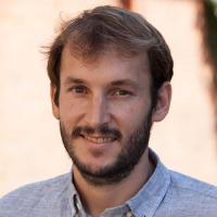
Dr. Roarke Horstmeyer is an assistant professor of biomedical engineering, electrical and computer engineering, and physics at Duke University. He develops microscopes, cameras and computer algorithms for a wide range of applications, from forming 3D reconstructions of organisms to detecting blood flow and neuronal activity deep within tissue. Most recently, Dr. Horstmeyer was a visiting professor at the University of Erlangen in Germany and an Einstein International Postdoctoral Fellow at Charitè Medical School in Berlin. Prior to his time in Germany, Dr. Horstmeyer earned a PhD from Caltech’s EE department (2016), an MS from the MIT Media Lab (2011), and bachelor’s degrees in Physics and Japanese from Duke in 2006.

